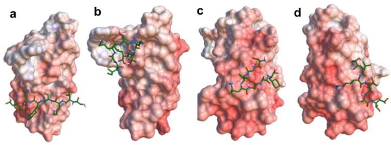
Figure 1.
Electrostatic surface of EDB fragment and 3D stick molecular models of linear peptides GVK (a), IGK (b), SGV (c), and ZD2 (d) fitted to the protein. For the surface of EDB, blue indicates positive charged residues, red represents negative areas and white are neutral regions. Active residues for docking calculations are numbered. For the peptide stick models, green indicates the carbon atoms, blue indicates the nitrogen atoms, white indicates the hydrogen atoms and red indicates the oxygen atoms.

