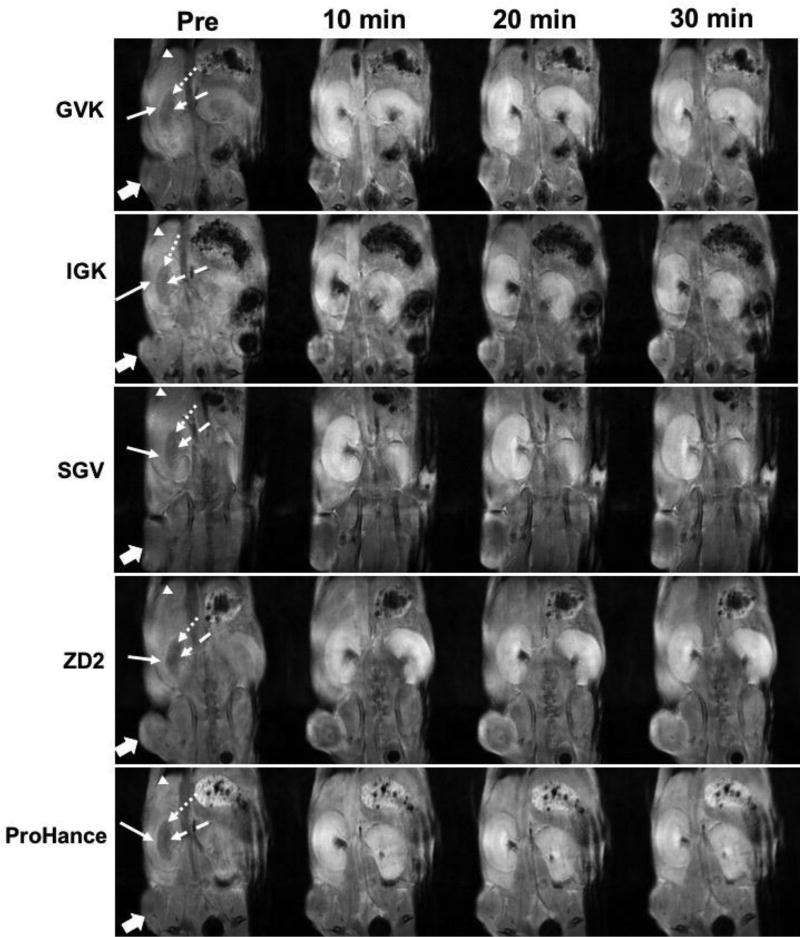
Figure 7.
T1-weighted 3D FLASH coronal MR images of PC3 tumor-bearing mice acquired pre-contrast (Pre) and at 10, 20 and 30 min after i.v. injection of GVK (a), IGK(b), SGV(c), ZD2(d) Gd-DOTA conjugates and Gd(HP-DO3A) (e) at 0.1 mmol/kg respectively. In the kidney cortex (full arrow) and outer medulla (round dot arrow), significant signal enhancement was found at 10, 20 and 30 min following injection of all peptide agents. In the inner medulla (dash arrow), high signal enhancement was found at 20 and 30 min as compared to at 10 min for the agents. In the liver, slight increase of signal (arrow head) at 10, 20 and 30 min after injection of the agents; In the tumor (block full arrow), all targeted agents produced significantly high tumor contrast enhancement as compared to the contrast agent Gd(HP-DO3A) (n = 5).

