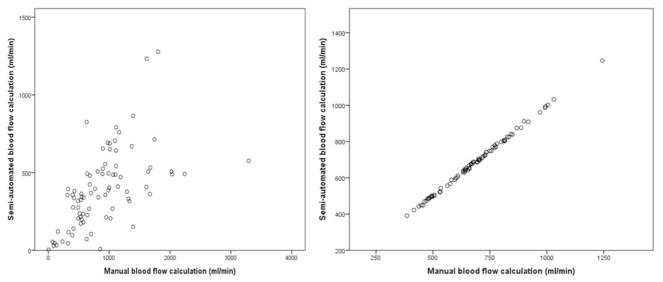
Figure 2.
Scatter plot showing the difference between the semi-automated and manual calculation of the venous blood flow (left) versus the arterial blood flow (right)
For representation of the venous blood flow volume measurement, the right upper IJV was used. The blood flow volumes represented on both axis are depicted as ml/min. For the corresponding arterial blood flow representation, the right upper CCA was used. IJV – internal jugular vein, CCA – common carotid artery.

