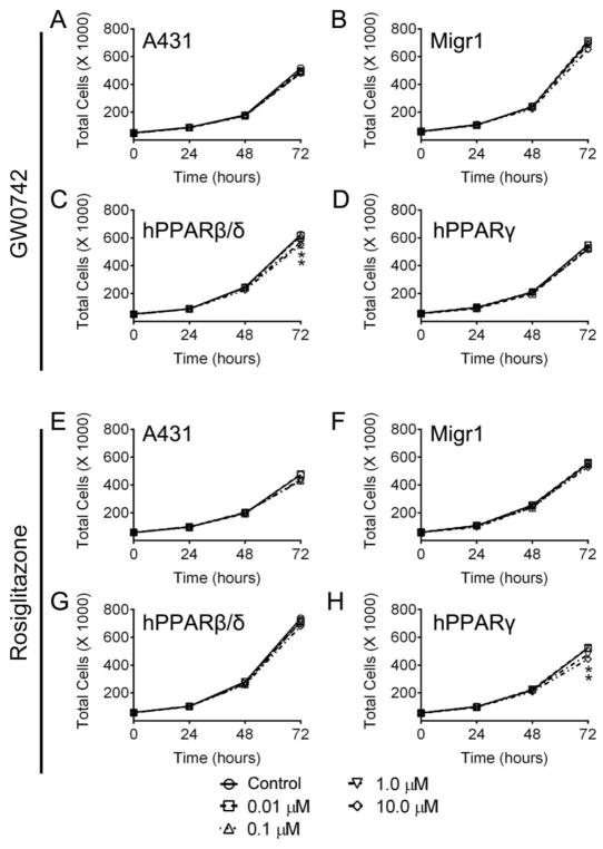
Fig. 3.
Effect of PPARβ/δ and PPARγ over-expression and/or ligand activation on cell proliferation. Proliferation of A431, A431-Migr1 vector control cells (Migr1), A431-Migr1-hPPARβ/δ cells (hPPARβ/δ), or A431-Migr1-hPPARγ cells (hPPARγ) was examined over a 72 h period by Coulter Counting. Cells were treated with indicated concentration of the PPARβ/δ ligand GW0742 (A–D) or the PPARγ ligand rosiglitazone (E–H), at time 0. Data represents triplicate independent sample means ± S.E.M.. *Significantly different than cell line-specific control (p ≤ 0.05).

