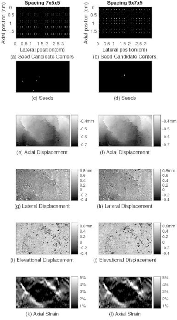
Fig. 9.

Motion tracking results for the same image plane shown in Fig. 5 through the in vivo human breast comparing displacement estimates obtained by the 3D RGMT with the spacing of seed candidate centers being 7×5×5 (left column) and 9×7×5 (right column). Comparing the images in the two columns, although the locations of the seed candidate centers and trusted seeds from which 3D RGMT start are different, similar motion tracking results are obtained, suggesting that the 3D RGMT is less dependent on the seed locations.
