Figure 1.
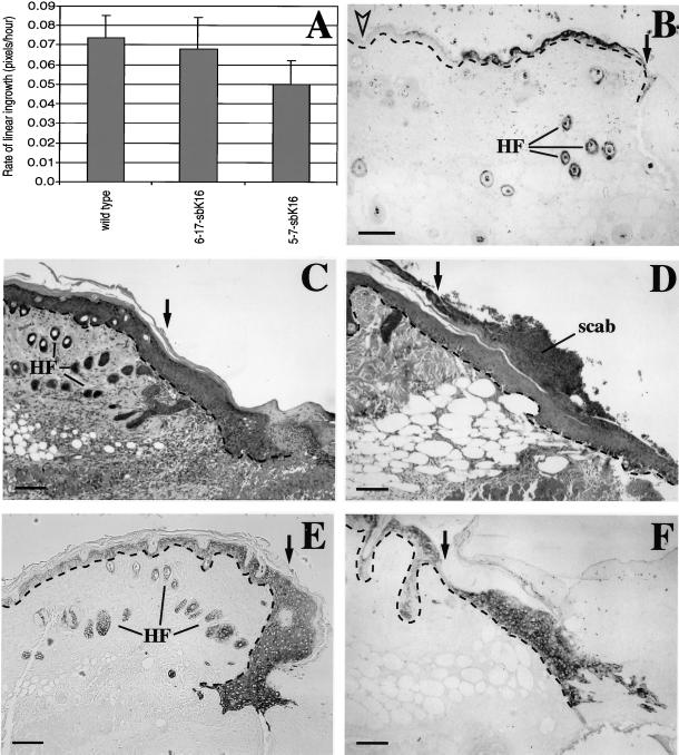
Analysis of full-thickness skin wounds in vivo. (A) In vivo wound closure assay was performed to determine the average rate of linear ingrowth for wild-type (n = 4), 6-17-sbK16 (n = 3), and 5-7-sbK16 (n = 3) mice. Mice from the 5-7-sbK16 line display significantly (p < 0.04) slower ingrowth than wild type. The 6-17-sbK16 mice do not vary significantly from wild type. (B–F) Skin wounds were fixed at various time points for histological analysis. C and E show wounds from wild-type mice, whereas B, D, and F show wounds from 5-7-sbK16 mice. Immunostaining with antibodies that preferentially recognize human K16 shows transgene induction 27 h after wounding (B). C and D are hematoxylin and eosin stainings of sections from 5-d-old wounds. E and F are 5-d-old wounds that have been immunostained with antibodies directed against K17. Black arrows denote the wound edge. The open arrowhead shows nonwounded skin, which does not express the transgene (B). HF, hair follicles. Bars, 100 μm.

