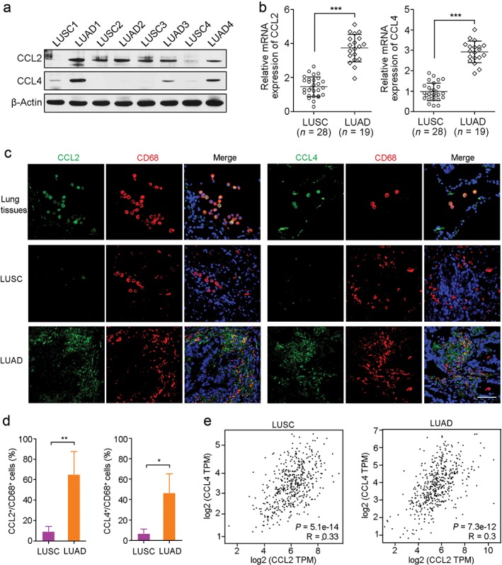
Figure 3.

CCL2 and CCL4 overexpression in lung adenocarcinoma (LUAD). (a) Western blot analysis showed that CCL2 and CCL4 were upregulated in LUAD but not in lung squamous cell carcinoma (LUSC). β‐Actin was used as a loading control. (b) Quantitative real‐time‐PCR showed high expression of CCL2 and CCL4 at RNA level in LUAD (n = 19) compared to LUSC (n = 28). ***P < 0.001. (c) Co‐staining of CCL2 or CCL4 (green) with macrophage marker CD68 (red) in normal lung, LUSC, and LUAD tissues. Scale bar, 100 μm. Cell nuclei were counterstained with 4′,6‐diamidino‐2‐phenylindole (blue). (d) The percentages of CCL2+/CD68+ and CCL4+/CD68+ cells per visual field in LUAD and LUSC tissues were analyzed. *P < 0.05. **P < 0.01. (e) Correlation analyses of CCL2 and CCL4 expression between LUSC and LUAD. mRNA, messenger RNA.
