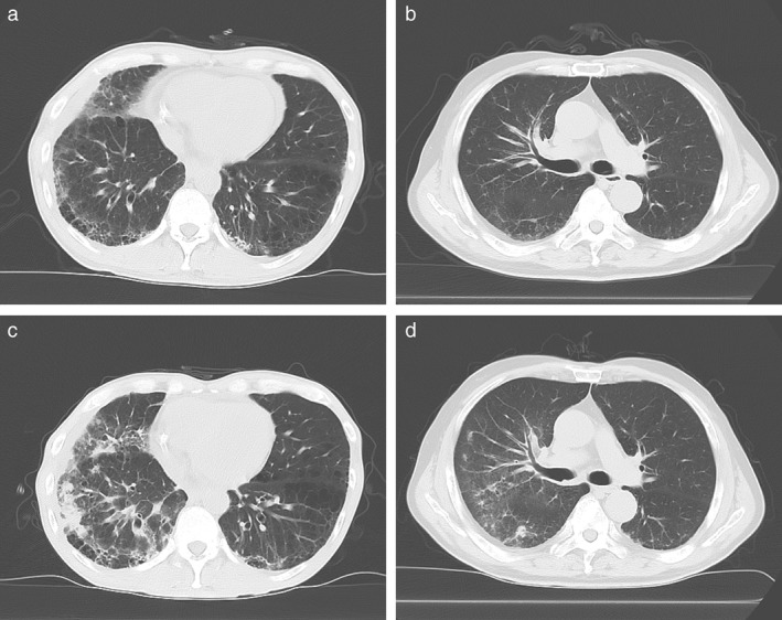
Figure 3.

The computed tomography (CT) images of the patient's lung were acquired (a,b) before the initial administration of nivolumab and (c,d) at the onset of nivolumab‐related ILD. (a,c) An example of the exacerbation type (Case 7 in Table 3), in which new ground glass opacities and consolidation are distributed along the site of preexisting ILD. (b,d) An example of the de novo type (Case 6 in Table 3), in which ground glass appearance and consolidation are distributed around lung metastases that are not involved with the preexisting ILD.
