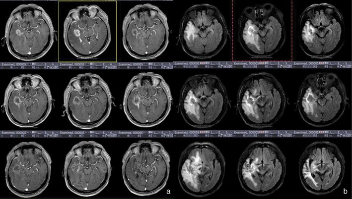
Figure 7.

Group 3 patients had a survival period of ≥ 30 months and were confirmed with tumor necrosis by surgical pathology. (a) Enhanced T1 weighted imaging shows increased volume and diffusion of lesions after radiotherapy. (b) T2 fluid‐attenuated inversion recovery (FLAIR) imaging shows a high‐signal ring sign at the periphery of the lesion.
