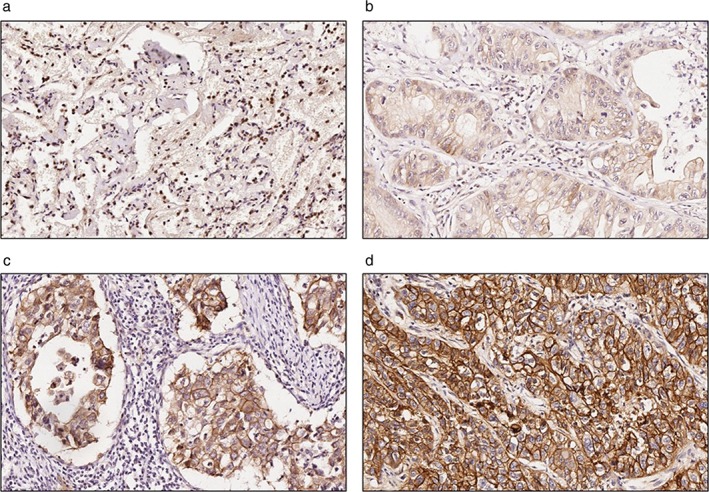
Figure 2.

Representative images of immunohistochemical (IHC) staining of S100A14 in normal or lung adenocarcinoma tissues. Faint or no signals of S100A14 were detected in normal lung specimens, whereas S100A14 was mainly expressed in the cell membrane and cytoplasm in lung adenocarcinoma tissues. (a) Normal lung tissue, (b) weak positive staining (IHC = 1), (c) positive staining (IHC = 2), and (d) strong positive staining (IHC = 3) in lung adenocarcinoma tissue.
