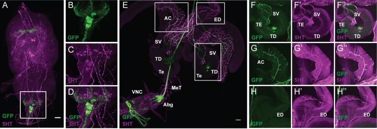
FIGURE 3.
FLP335 restricted expression of fru-GAL4 with tsh-GAL80 targeted a subset of abdominal serotonergic fru neurons. (A–D) Double staining of male VNC with anti-GFP (green) and anti-5HT (magenta) of a Z-stack projection. (E) Double staining of the male reproductive organs with anti-GFP (green) and anti-5HT (magenta). (F–H) Co-localization of GFP and 5HT signal at the seminal vesicle (F), the accessory glands (G), and the absence of GFP signal at the ejaculation duct (H). AC, accessory gland; ED, ejaculation duct; SV, seminal vesicle; TD, testicular duct; Te, testes; MeT, median trunk nerve; VNC, ventral nerve cord; Abg, abdominal ganglion. Scale Bar = 50 μm. The brightness of E had been adjusted in ImageJ so that the innervation at the peripheral tissues can be visualized [B: Mean Intensity in Red/Blue Channel (displayed as magenta) = 38.06 ± 41.28, Mean Intensity in Green Channel = 6.63 ± 15.75; E: Mean Intensity in Red/Blue Channel (displayed as magenta) = 20.53 ± 31.66, Mean Intensity in Green Channel = 11.62 ± 24.89].

