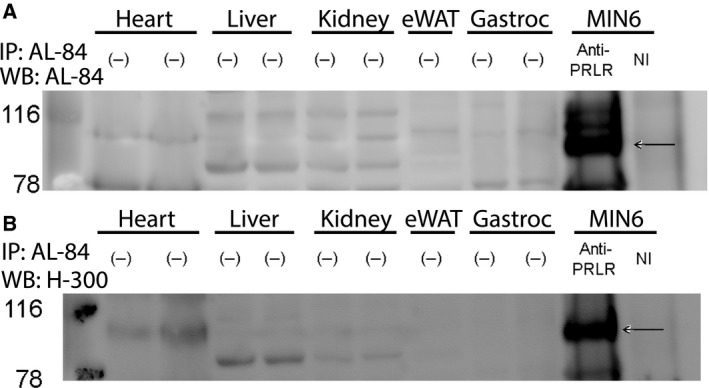
Figure 4.

PRLR immunoblot of tissue lysates from various mouse tissues and in MIN6 mouse insulinoma cells (positive control). The black arrows denote the PRLR in the IP control. (−) denotes that no immunoprecipitation was performed. NI denotes nonimmune serum. Immunoprecipitation was performed with anti‐PRLRcytAL‐84, and resolved eluates were immunoblotted sequentially with (A) anti‐PRLRcytAL‐84 and (B) anti‐PRL‐R (H‐300).
