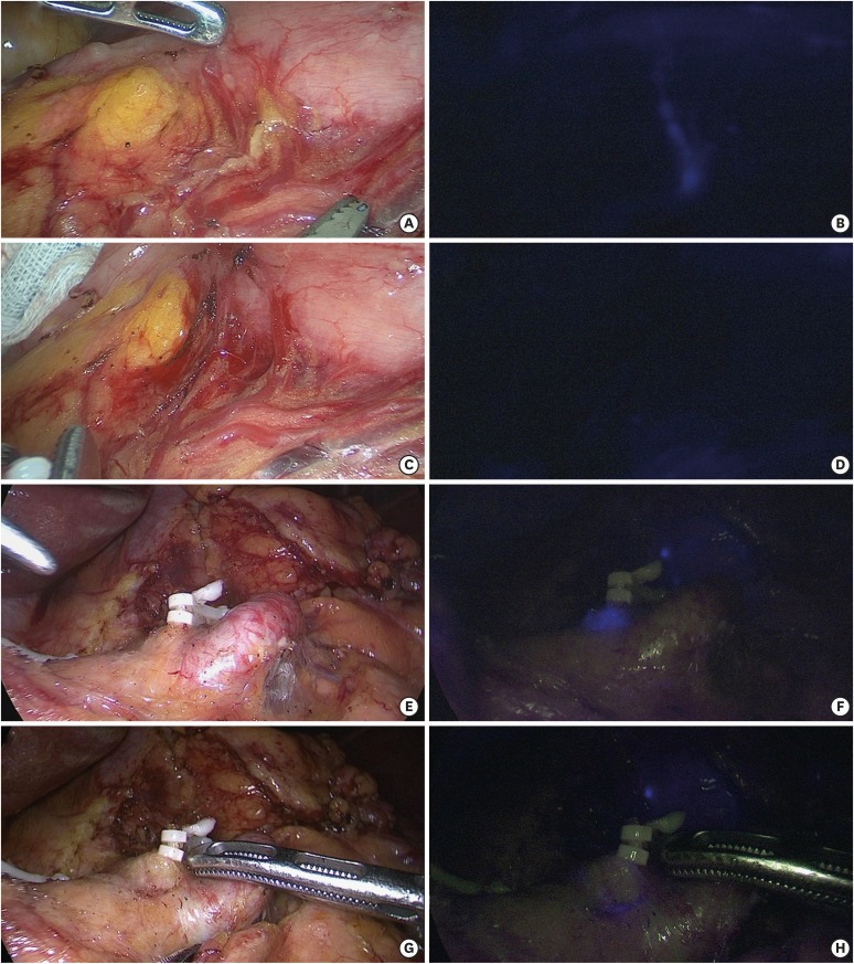
Fig. 3. Comparison of additional tissue dissection guided with a NIR camera. Before dissection of the near infra-artery in a case of LPPG: (A) under white light view and (B) in NIR mode. After dissection: (C) under white light view and (D) confirmation with the NIR mode. After dissection, tissue with additional ICG uptake (E-tissue) in the left gastric artery in LDG: (E) under white light and (F) in NIR mode. After additional dissection of the E-tissue (G) under white light and (H) in NIR mode.
NIR = near-infrared; LPPG = laparoscopic pylorus-preserving gastrectomy; ICG = indocyanine green; LDG = laparoscopic distal gastrectomy.

