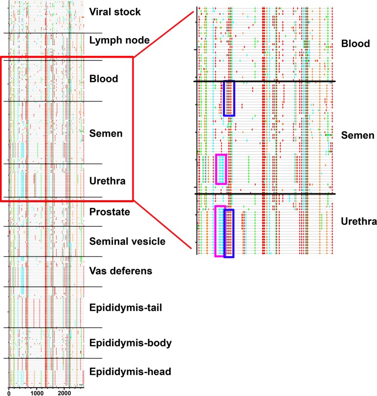
FIG 8.
Comparison of semen and urethra sequence motifs in macaque H2. (Left) Highlighter plots generated from all sequences derived from macaque H2 together with viral stock. Sequences are grouped by tissue. (Right) Detailed view of the area indicated on the left within the red rectangle. The two 150-bp motifs which are absent from any blood related sequences are observed either separated in semen-related sequences or fused in urethra sequences. This pattern may result from recombination events between semen sequences after infection of the urethra.

