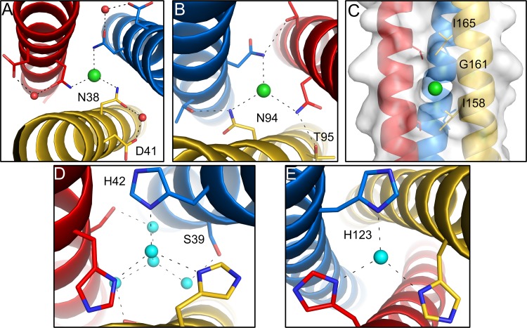
FIG 3.
Chloride ions and water molecules bound in the core of the σ1 coiled coil. The trimeric σ1 protein is shown in blue, red, and yellow. Chloride ions are shown as green spheres, and water molecules are shown as cyan spheres. (A and B) Enlarged views of T1L σ1 showing bound chloride ions complexed with N38 (A) and N94 (B), respectively. (C) A third chloride ion is bound in a hydrophobic pocket inside the core of the structure. (D and E) Enlarged views of T3D σ1 showing bound water molecules in the coiled-coil interior that form hydrogen bonds with H42 (D) and H123 (E).

