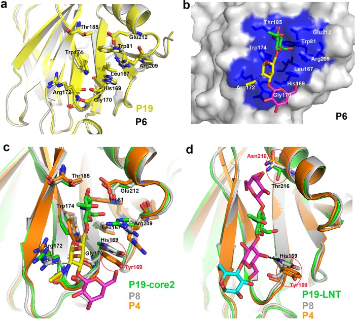
FIG 5.
Structural characteristics of the glycan binding site in P[6]/P[4]/P[8] VP8*s. (a) Comparison of the conformations of the residues involved in glycan binding in P[19] (PDB ID 5GJ6; yellow) and that of porcine P[6] VP8* structure (PDB ID 5YMU; gray). The amino acids involved in the interaction are shown in stick representation. (b) Superimposition of core 2 on the putative binding site of porcine P[6] VP8* structure (PDB ID 5YMU; gray). P[6] VP8* is shown in surface representation, and the putative binding site is colored blue. (c) Structure superimposition of P[8] and P[4] VP8*s on P[19] VP8*-core 2. Core 2 is shown as sticks (GlcNAc, green; GalNAc, yellow; Gal, magenta;). Residues involved in the core 2 binding are shown as sticks. Tyr169 in P[4]/P[8] is circled and colored red. VP8*s of P[19], P[8], and P[4] are colored green, gray, and orange, respectively. (d) Structure superimposition of P[8] and P[4] VP8*s on P[19] VP8*-LNT. LNT is shown as sticks (Gal, magenta; GlcNAc, green; Glc, cyan). Residues 169 and 216 are shown as sticks. The hydrogen bond between His169 and LNT is shown as a black dotted line. Tyr169/Asn216 in P[4]/P[8] are circled and colored red. P[19], P[8], and P[4] are colored as in panel c.

