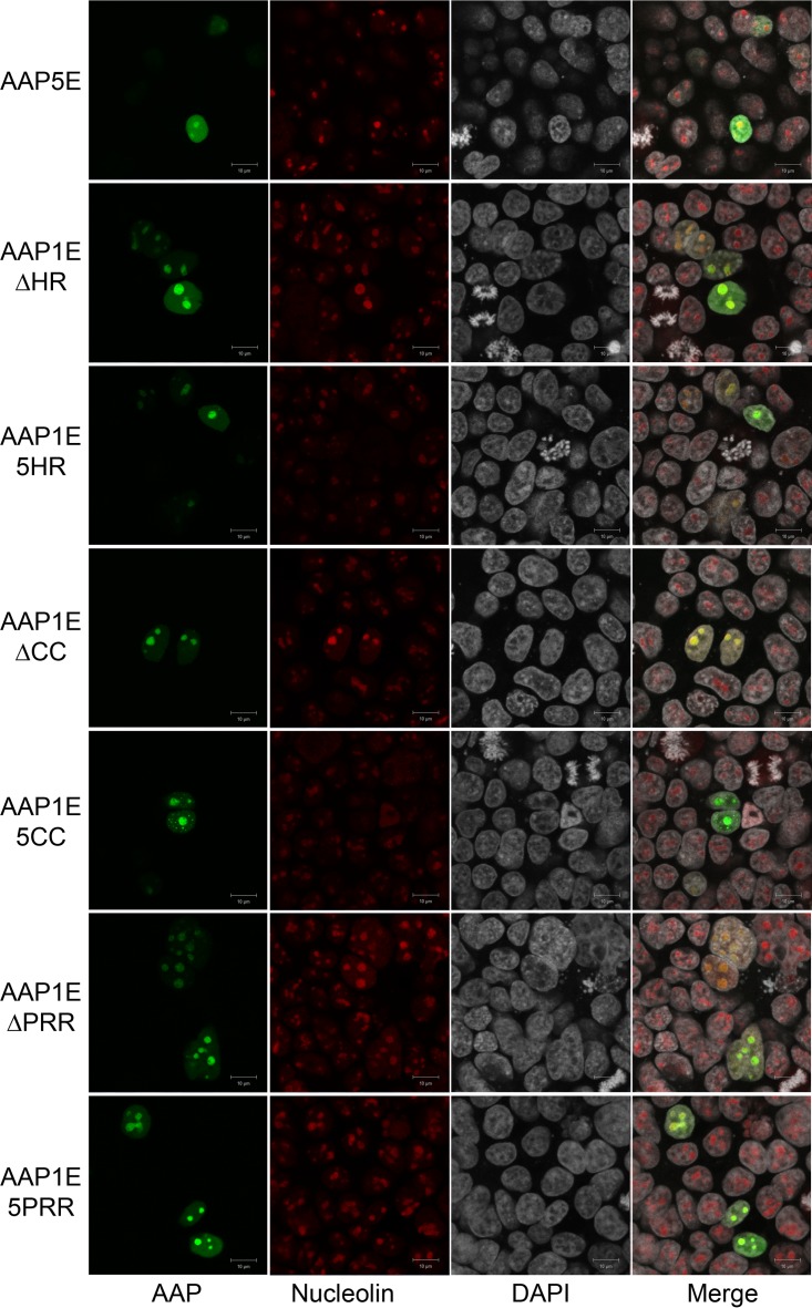
FIG 6.
Confocal microscopy of different AAP5E and AAP1E derivatives. AAP5E and AAP1E derivatives were transfected as described in Materials and Methods. AAP5E and AAP1E derivatives were visualized by their native EGFP fluorescence, nucleoli were immunostained with C23 antibody, and nuclei were stained with DAPI.

