FIG 7.
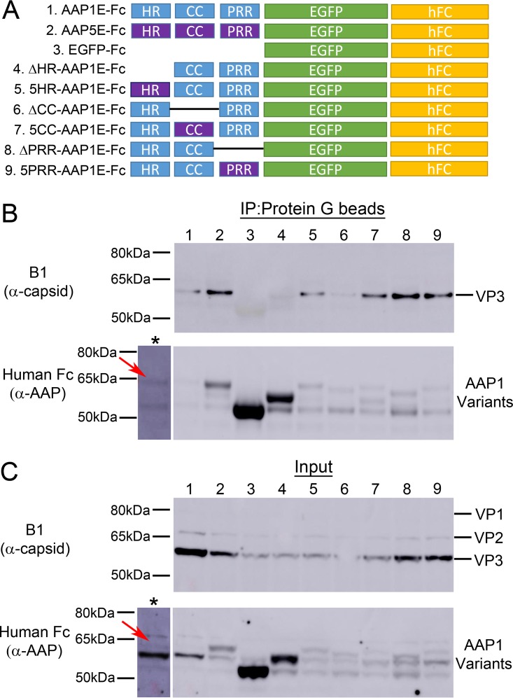
Immunoprecipitation of VPs by different N-terminal mutants of AAP1. (A) Schematic of different AAP mutants. The N-terminal domains are either deleted or replaced with AAP5 counterparts (purple). All AAP constructs have their BRs replaced by the human Fc domain at the C terminus. (B and C) Representative Western blot images of immunoprecipitation (B) and input (C) of VPs by AAP1E mutants. The asterisk (*) lane represents an overexposure of lane 1, and red arrows denote the position of AAP1E-hFc. Samples were collected at 3 days posttransfection of HEK293 cells with pXX680, pXR1ΔAAP1, pCDNA3.1-AAP1E-Fc, and pTR-CBA-Luc. Detailed protocols are described in Materials and Methods. AAP1E-Fc was pulled down using magnetic protein G beads and is immunostained with goat α-human-HRP antibody; VP is stained by mouse α-capsid antibody (B1).

