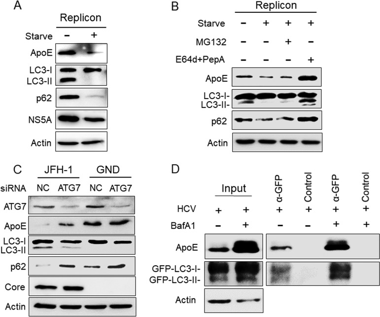
FIG 4.
Analysis of the effect of autophagy on ApoE. (A) HCV GLR replicon cells without treatment or nutrient starved in HBSS for 5 h were lysed for immunoblot analysis. (B) GLR cells were not treated or were treated with HBSS for 5 h in either the absence or presence of 10 μM MG132 or 25 μM E64d and 50 μM pepstatin A (PepA). Cells were then harvested for immunoblot analysis. (C) Huh7 cells were electroporated with JFH-1 or GND RNA and 24 h later were transfected with the control (NC) siRNA or the ATG7 siRNA. Cells were harvested at 48 h after the siRNA transfection for immunoblot analysis. (D) Huh7-GFP-LC3 cells were infected with HCV (MOI = 1) and treated with either DMSO or BafA1. GFP-LC3 and its associated autophagosomes were immunoprecipitated with control beads or anti-GFP antibody-conjugated beads as described in Materials and Methods, followed by immunoblot analysis using the anti-ApoE and anti-GFP antibodies. Total cell lysates were also analyzed by immunoblotting to serve as the input control.

