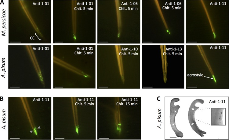
FIG 1.
Stylin-01 is present in the acrostyle. (A) Immunolabeling of M. persicae (top) and A. pisum (bottom) dissected maxillary stylets untreated or predigested with chitinase (Chit.). Fluorescence signals were more or less intense depending on the antibody used and visible at the tip of maxillary stylets in the common canal (CC) and in the acrostyle. The antibodies for which no labeling has been observed are not shown (anti-08 and anti-09). (B) Anti-1-11 antibody directly labeled the surface of the acrostyle of aphid maxillary stylets and was weaker following a 5- to 15-min chitinase digestion treatment. (C) Transmission electron microscopy (TEM) observations of cross sections of the upper part of the common canal of A. pisum (indicated by gray arrowheads on the leftmost image of panel B). The darker density at the surface of the inner cuticle corresponds to the acrostyle (white arrows). Immunogold labeling with anti-1-11 antibody (right) showed gold particles at the surface of the acrostyle (magnified in the inset). Scale bars represent 5 μm in all immunofluorescence images, 100 nm in the TEM panel, and 50 nm in the inset.

