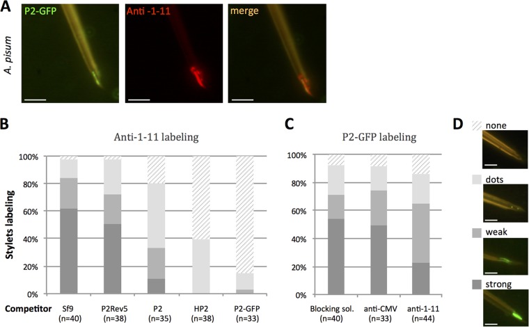
FIG 4.
CaMV protein P2 and anti-1-11 IgGs colocalize in and compete for the acrostyle. (A) Coincubation of P2-GFP and anti-1-11 antibody with A. pisum dissected stylets. P2-GFP (green fluorescence) and anti-1-11 antibody (red fluorescence) colocalize on the acrostyle (seen as orange labeling). (B) Histograms show the proportion of maxillary stylets with the acrostyle labeled by anti-1-11 antibody from 4 independent experiments, after preincubation with crude extracts from healthy Sf9 cells (Sf9), from Sf9 cells producing P2, P2Rev5 mutant, P2-GFP fusions, or purified His tag-P2 (HP2), as indicated. While no significant difference was observed in the intensity of anti-1-11 labeling after Sf9 or P2rev5 preincubation (one-way ANOVA, P = 0.779), the proportion of stylets strongly labeled was significantly reduced when P2, P2-GFP, and HP2 were used as competitors (one-way ANOVA, P = 2.27 × 10⁻⁵). (C) Histograms show the proportion of maxillary stylets with the acrostyle labeled by P2-GFP from 4 independent experiments, after preincubation with blocking solution containing no antibody, anti-CMV as a negative control, and anti-1-11 antibodies. The intensity of the labeling observed after preincubation with blocking solution containing anti-1-11 antibodies is reduced compared to that with stylets preincubated with blocking solution or anti-CMV as a negative control. However, this reduction is not significant (ANOVA, P = 0.0503). The numbers of maxillary stylets observed for each treatment is indicated as “n.” Each labeled stylet counted in panels B and C was scored as strongly, weakly, dot, or not labeled; a representative image illustrating each of these scores is shown in panel D. Scale bars represent 5 μm.

