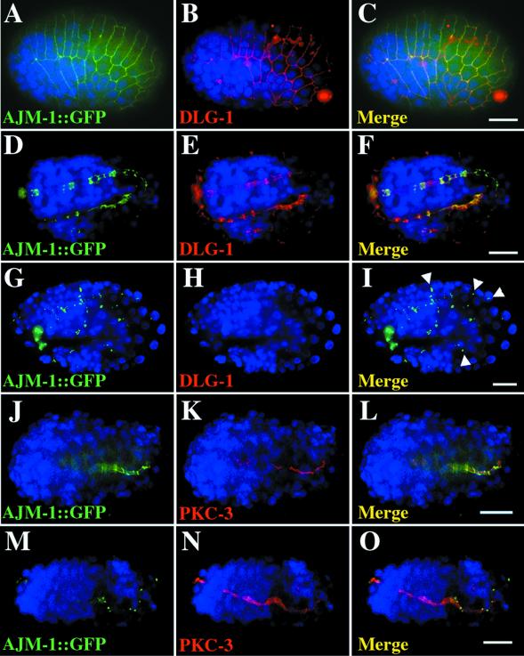
Figure 2.
DLG-1 is localized to adherens junctions and required for AJM-1:: GFP localization. Immunostaining of embryos that express AJM-1:: GFP (green, A, D, G, J, M) with anti-PSD-95 antibodies (red, B, E, H) or with anti-PKC-3 antibodies (red, K, N). Nuclei are stained with DAPI (blue). Merged images (C, F, I, L, O) contain a 10 μm scale bar. AJM-1:: GFP and DLG-1 colocalize to adherens junction on the epidermis (A-C, lima bean stage) and in the intestine (D-F, tadpole stage) of wild-type embryos. (G-I, twofold stage) AJM-1:: GFP is found in small punctate structures between cells in dlg-1(RNAi) embryos No DLG-1 protein is detected in these embryos. (J-L, gastrulating embryo) AJM-1:: GFP is found at intestinal adherens junctions (green) whereas PKC-3 is localized to the apical surface of these cells (red) in wild-type embryos. (M-O, gastrulating embryo) AJM-1:: GFP is found in punctate structures, but PKC-3 is found at the apical surface of cells from dlg-1(RNAi) embryos.

