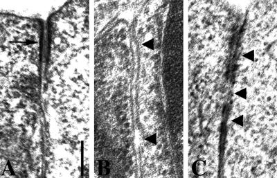
Figure 3.
Abnormal ultrastructure of adherens junctions in dlg-1(RNAi) embryos. (A) Electron microscopy of an adherens junction (arrow) in a wild-type embryo. (B) Electron-dense adherens junction material is often missing in dlg-1(RNAi) embryos. Arrowheads point to gaps that have formed between adjacent membranes. (C) When adherens junctions are visible in dlg-1(RNAi) embryos, they are often discontinuous (arrowheads). Scale bar is 200 nm.

