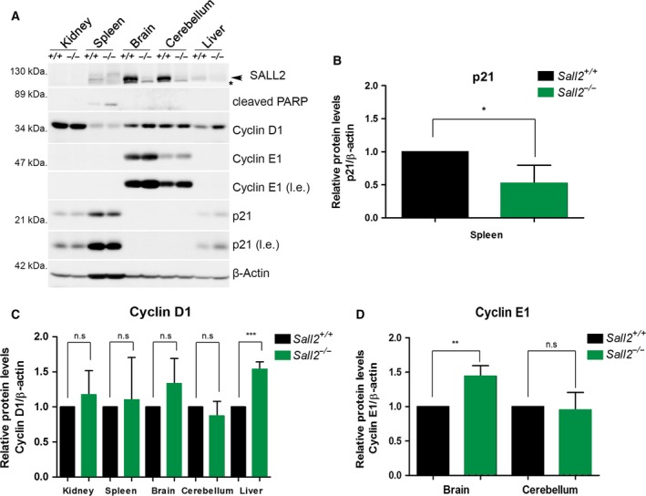
Figure 4.

Increased expression of cyclins D1/E1 in tissues from Sall2‐deficient mice. Tissues from 6‐ to 8‐week‐old Sall2 +/+ and Sall2 −/− mice were isolated and lysed to evaluate SALL2, cyclin D1 and cyclin E1 levels by western blot analysis (A). Representative western blot of tissues analyzed. An arrow indicates SALL2, and the asterisk corresponds to nonspecific band. p21—a protein positively regulated by SALL2 (Li et al., 2004)—was used as positive control; β‐actin was used as normalizer. l.e; long exposure. (B–D) Densitometric data from western blots of p21, cyclins D1 and E1 from five isogenic mouse/genotype. Data are expressed as mean ± SD from five independent mouse tissues per genotype. *P ≤ 0.05, **P ≤ 0.01, ***P ≤ 0.001, Student′s t–test; n.s, nonsignificant.
