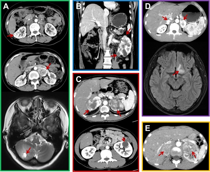
Figure 1.
Representative CT and MRI scans from the probands. (A) CT and MRI scans showing lesions in proband 1, including a right renal cell carcinoma (RCC) (upper image) and left pheochromocytoma (Pheo) (middle image) by CT scan, and a hemangioma (HB) in the right cerebellum (bottom image) by MRI scan. (B) A CT scan showing RCCs in proband 2. (C) CT scans of Pheos (upper image) and a renal cyst (bottom image) in proband 3. (D) CT and MRI scans showing the Pheos (upper image) and HB (bottom image) in proband 4. (E) CT scans showing Pheos in proband 5. Red arrows indicate the relevant features.

 This work is licensed under a