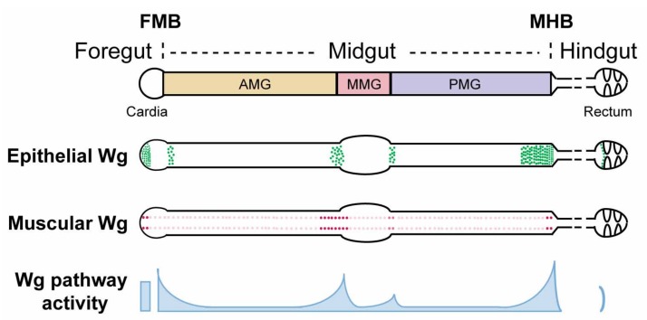
Figure 1.
Schematic view of wg expression and Wg pathway activation in the Drosophila adult gut. The Drosophila adult gut is divided into foregut, midgut, and hindgut. The foregut/midgut boundary (FMB) and midgut/hindgut boundary (MHB) provide local niches for region-specific stem cells and contain critical valves that regulate food entry and exit. The midgut is further partitioned into anterior midgut (AMG), middle midgut (MMG), and posterior midgut (PMG), based on major constrictions and the existence of a specific acid-secreting region in the MMG. A wg-gal4 knock-in line driving UAS-lacZ reveals wg expression in both the epithelium and the surrounding visceral muscle. At major compartment boundaries of the midgut, epithelial sources of wg are detected within enterocytes. In addition, four rows of wg-expressing cells are detected in the surrounding circular visceral muscles throughout the entire length of the midgut. Instead of being uniform, these muscle sources of wg are enriched at major compartment boundaries. Similarly, Wg pathway activation exists in gradients, exhibiting high-level expression at compartment boundaries and low-level expression throughout compartments.

