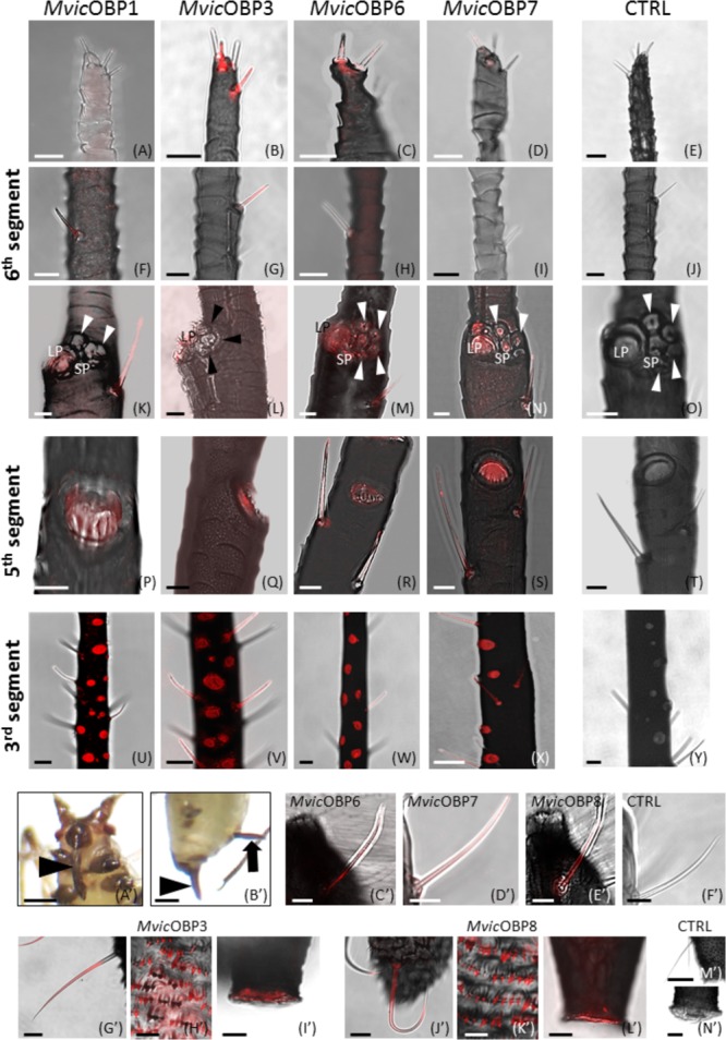
FIGURE 4.

(A–Y) Whole-mount immunolocalization experiments showing the OBP expression in type II trichoid sensilla located on the antennal tip (A–D), in type II trichoid sensilla on the 6th antennal segment (F–I), in primary rhinaria on the 5th (K–N) and 6th segments (P–S) and in secondary placoid sensilla on the 3rd segment (U–X). (E,J,O,T,Y) Negative controls. Bars in (A–T), 10 μm; bars in (U–Y), 25 μm. (A’–N’) Whole-mount immunolocalization experiments showing the OBP localization in the mouthparts (arrowhead in (A’)), in the cauda (arrowhead in (B’)) and in cornicles (arrow in (B’)).(E–G,C’–E’) Immunolocalization of OBPs in the long sensilla on the labium sides. (G’–L’) OBPs detection in hair-like structures and finger-like projections in cauda and in cornicles. (F’,M’,N’) Negative controls. Bars in (A’,B’), 250 μm; bars in (C’–F’,H’,K’), 10 μm; bars in (M’), 50 μm; bars in (G’,I’,J’,L’,N’), 20 μm
