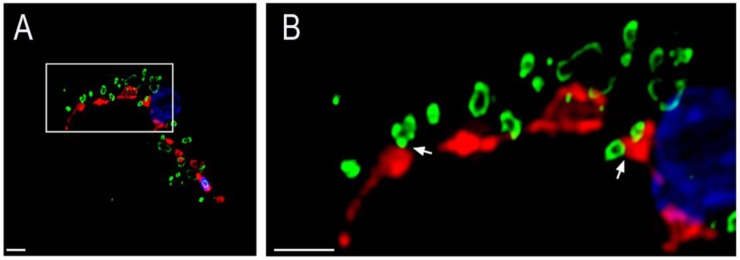
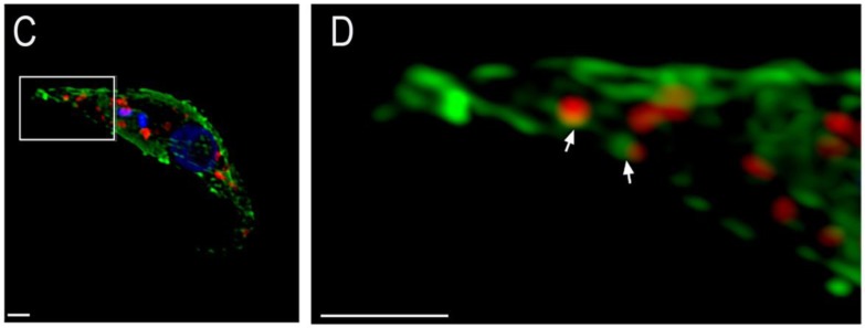
Figure 1.
Representative super resolution images of T. brucei acidocalcisomes and mitochondria in BSF (A,B) and procyclic form (PCF) (C,D) trypanosomes. Close proximity between the acidocalcisomes and mitochondria could be observed in both life cycle stages and are indicated by white arrows in the magnified images (B,D). Mitochondria are in red in A/B and in green in C/D. Acidocalcisomes are in green in A/B and in red in B/C. DAPI staining of nuclei and kinetoplasts is in blue. Scale bar = 1 µm.


