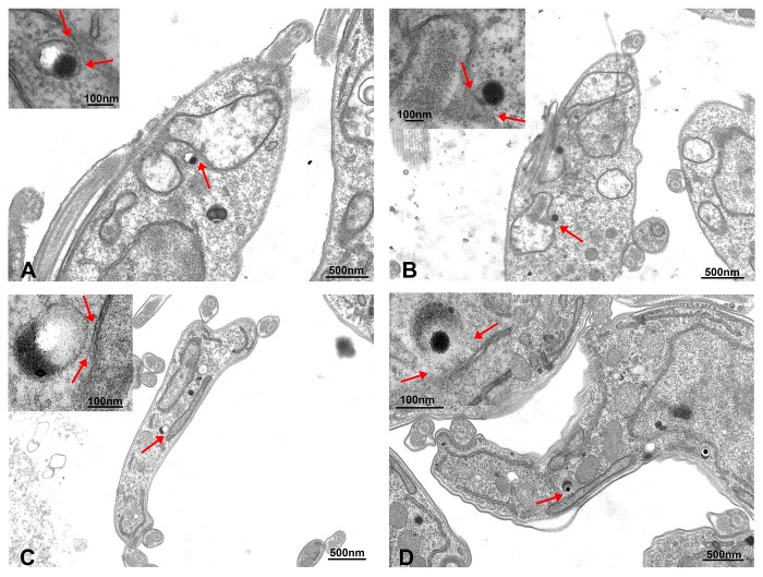
Figure 2.
Representative transmission electron microscopy images of PCF (A,B) and bloodstream form (BSF) (C,D) T. brucei showing contacts between acidocalcisomes and the mitochondrion of the parasites. Acidocalcisomes appear as rounded organelles containing electron-dense material that adheres to one side of the membrane, and are seen adjacent to the mitochondrion double membrane. The contact sites could be observed in both life cycle forms and are indicated by red arrows in the insets at higher magnification.

