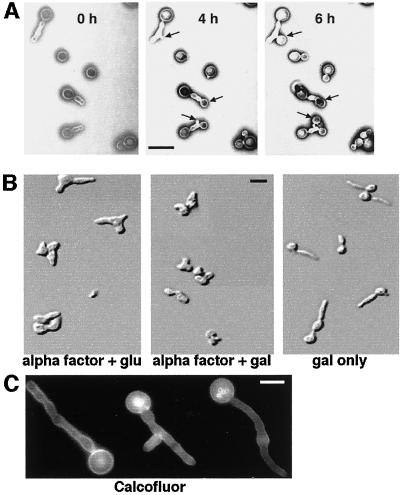
Figure 1.
Characterization of INT1-filaments in S. cerevisiae. (A) The filaments induced by INT1 expression are not in a terminal physiological state. YJB5763 was grown in galactose to induce INT1-filaments and then immobilized onto agar containing glucose to repress GAL1-INT1. Images were then obtained at the times indicated. Sites of new bud growth are indicated by arrows. Bar, 10 μm. (B) INT1-filaments do not form in α-factor–arrested cells. DIC micrographs of YJB5763 were arrested with α-factor as described in MATERIALS AND METHODS, washed once with sterile water, and resuspended in medium containing α-factor with glucose (glu) or galactose (gal), or galactose without α-factor, as indicated. Bar, 10 μm. (C) Chitin localization is diffuse in INT1-induced filaments. Shown is fluorescence micrograph of strain YJB5763 expressing INT1 and stained with Calcofluor white. Bar, 5 μm.

