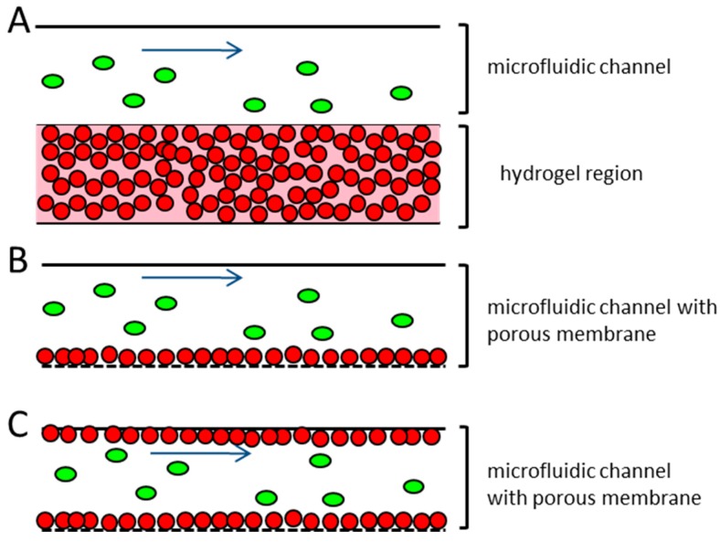
Figure 1.
Schematic representation of different microfluidic systems used for the research on the vessel associated steps of the metastatic cascade (A) endothelial cells are embedded in hydrogel and injected into the microfluidic channel, where they are arranged in a capillary network. Tumor cells are introduced to the top of the hydrogel, and can migrate into it. No perfusion of the tubule-like structures takes place; (B) Endothelial cells are seeded in a monolayer on top of a porous membrane. The confluency of the endothelial cells on the membrane can often not be guaranteed. The tumor cells are introduced to the system under flow conditions; (C) Proposed microfluidic device. Endothelial cells are seeded to the total dimension of the microfluidic channel and the adjacent porous membrane, representing the vessel equivalent. The confluency of the endothelial cells is confirmed before the experiment. Tumor cells are introduced to the device under different flow conditions. Red dots—endothelial cells; green ovals—tumor cells; blue arrow—direction of flow.

