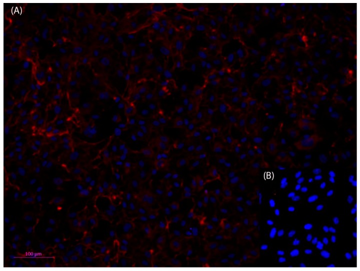
Figure 3.
Fluorescent microscopic photos of anti-Collagen IV, Cy3 (red) immunofluorescence staining of human primary pulmonary arterial endothelial cells (HPAEC) (A); nuclei stained with Hoechst dye (blue). Collagen IV can be detected both in the cytosol of the cells as well as outside of the cells, which indicates that the HPAEC cells have formed an adequate basement membrane (B). The negative control shows that the method has been used appropriately. Dilution of antibodies: 1st antibody anti collagen IV 1:50, and 2nd antibody Cy3 anti rabbit 1:500). Scale bar 100 µm.

