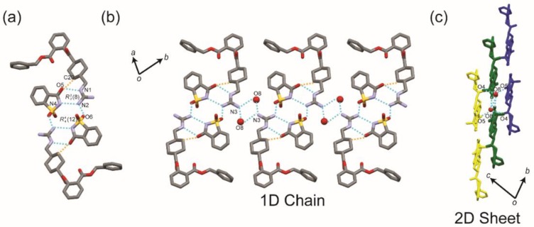
Figure 4.
The hydrogen bond architecture in the BEX–SAC crystal. (a) The interaction involving two sets of cationic benexate and anionic saccharinate molecules and water constructs. (b) 1D chain structures parallel to the (110) plane. Each 1D chain (represented by different colors) interacts with each other to form (c) a 2D sheet structure. Conventional and non-conventional hydrogen bonds are drawn by dashed blue and orange lines, respectively. Hydrogen atoms have been omitted for clarity. The oxygen atoms from water molecules are drawn in ball-setting.

