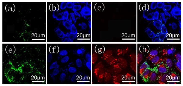
Figure 7.
Confocal microscopy images of HeLa cells incubated with 12.5 μg/mL (a–d) MIL-100(Al) gels and (e–h) DOX-loaded MOGs, respectively. Blue fluorescence represents the living cell imaging. Red fluorescence represents the released DOX from DOX-loaded MOGs within the cancer cells. Green fluorescence represents the apoptosis of cells. (d,h) are the merged images of (a–c) and (e–g), respectively.

