Figure 8.
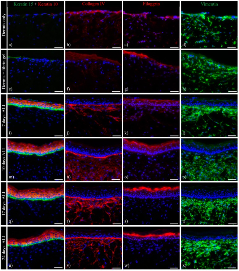
Immunohistochemistry of ftSEs. Expression of skin-specific markers was observed over time. Representative images of (a,d) dermis only and (e,f) dermis with added fibrin gel, as well as ftSEs after (i–l) seven days air lift (ALI), (m–p) 10 days ALI, (q–t) 17 days ALI, and (u–x) 24 days ALI were selected for further analysis. Column 1 (a,e,i,m,q,u) shows keratin 10 (red)/15 (green) double immunostaining. Column 2 (b,f,j,n,r,v) shows collagen IV, Column 3 (c,g,k,o,s,w) shows filaggrin, and Column 4 (d,h,l,p,t,x) shows vimentin staining. An increase in epidermal differentiation and basement membrane marker expressions at different time points indicates a physiologically relevant skin development. Counterstaining of cell nuclei with DAPI (blue). Scale bar: 50 µm.

