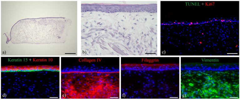
Figure 10.
HE staining and immuno-labeling of ftSE cultured for seven days in the 2OC. (a,b) HE stain of the ftSE showed preserved morphology comprising a multilayered epidermis. (c) TUNEL-Ki67 (green and red) staining of the ftSE revealed a considerable number of proliferating cells within the basal layer of the epidermis, while only a few apoptotic cells could be detected. (d) Keratin 10 (red)/15 double stain, (e) collagen IV (red), (f) filaggrin (red), and (g) vimentin (green) staining indicate advanced levels of differentiation within the ftSE cultured in the 2OC. Counterstaining of cell nuclei with DAPI (blue). (a) Scale bar: 500 µm. (b,d–g) Scale bar: 50 µm. (c) Scale bar: 100 µm.

