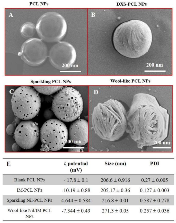
Figure 1.
Scanning electron microscopy (SEM) images of blank PCL NPs (A), IM-DXS loaded PCL NPs (B), burst sparkling Nil loaded PCL NPs (C) incubated in acid condition (pH 5.0) and wool-like hollow Nil/IM loaded PCL NPs (D). Magnification: 34.01 KX. Scale bars: 200 nm. Physicochemical characterization of different PCL NPs formulation (E). Representative measurements of three distinct sets of data have been reported (Student’s t-test, P-values < 0.05).

