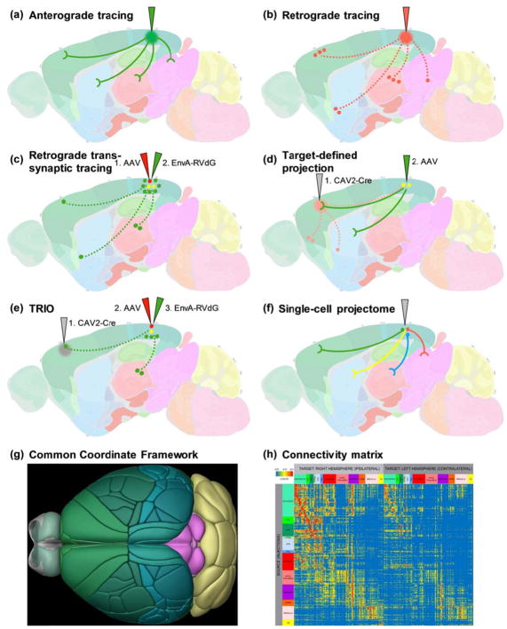
Figure 1.
Representative approaches for mesoscale connectomics. (a) In anterograde tracing, traditional or viral anterograde tracer is injected in the source region of interest and tracer-labeled long-range axonal projections and terminals in other parts of the brain are mapped. (b) In retrograde tracing, traditional or viral retrograde tracer is injected in the target region of interest and tracer-labeled somata or nuclei of input neurons in other parts of the brain are mapped. Dotted lines indicate that axon fibers of input neurons may or may not be labeled by the tracer. (c) In the most common scenario of monosynaptic, retrograde trans-synaptic tracing, AAV helper virus(es) (red) expressing TVA and RV-G proteins under Cre control is first injected into the target region of interest of a Cre-driver-containing animal, and then EnvA-pseudotyped, G-deleted rabies virus (RVdG) (green) is injected into the same region. Only neurons infected with both AAV and rabies virus (called starter cells, which are turned from red to yellow) will produce rabies viral particles that will be transported across synapses and label the somata of presynaptic input neurons (green cells) both locally and in the long distance. (d) In target-defined projection mapping, CAV2-Cre (or another retrograde virus, gray) is first injected into a target region of interest, and it will be taken up retrogradely by neurons projecting to the target region (pale red paths and cells); then a Cre-dependent AAV is injected into one of the source regions of interest, and it will label only the neurons (yellow) projecting to the initial target region as they contain CAV2-derived Cre. The axonal projections of these target-defined neurons will then be visualized (green fibers). Pale red indicates that CAV2-Cre infected cells may or may not be visualized, depending on whether the animal contains a Cre-reporter or not. (e) In TRIO (tracing the relationship between input and output), two steps of retrograde tracing are involved. CAV2-Cre (or another retrograde virus, gray) is first injected into a target region of interest, and it will be taken up retrogradely by neurons projecting to the target region. Then a (set of) Cre-dependent AAV helper virus(es) is injected into one of the source regions of interest, to express TVA and RV-G proteins only in the neurons (red) projecting to the initial target region as they contain CAV2-derived Cre. Finally, EnvA-pseudotyped, G-deleted rabies virus (RVdG) (green) is injected into the same source region. Only neurons infected with CAV2-Cre, AAV and rabies virus (note these are target-defined starter cells, which are turned from red to yellow) will produce rabies viral particles that will be transported across synapses and label the somata of presynaptic input neurons (green cells) both locally and in the long distance. (f) In single-cell projectome, individual neurons (green, red and blue) in the source region of interest are sparsely and strongly labeled by transgenic or viral expression of a fluorescent protein, and their full extent of dendritic and axonal arbors are imaged and reconstructed to reveal each neuron’s unique set of projection targets (green, red and blue fibers). Sometimes two neurons with otherwise different targets may also share one or more common target(s) (as indicated by the yellow fiber). Alternatively, individual neurons can also be densely labeled by barcoded anterograde viruses, and their individual projection targets resolved through the barcodes. (g) The mouse brain Common Coordinate Framework (CCF) is a digital 3-D reference atlas for image data registration and anatomical delineation. (h) Segmented and quantified mesoscale connectomic datasets can be used to generate connectivity matrix for neural network analysis (adapted from Ref [53]).

