
Figure 5.
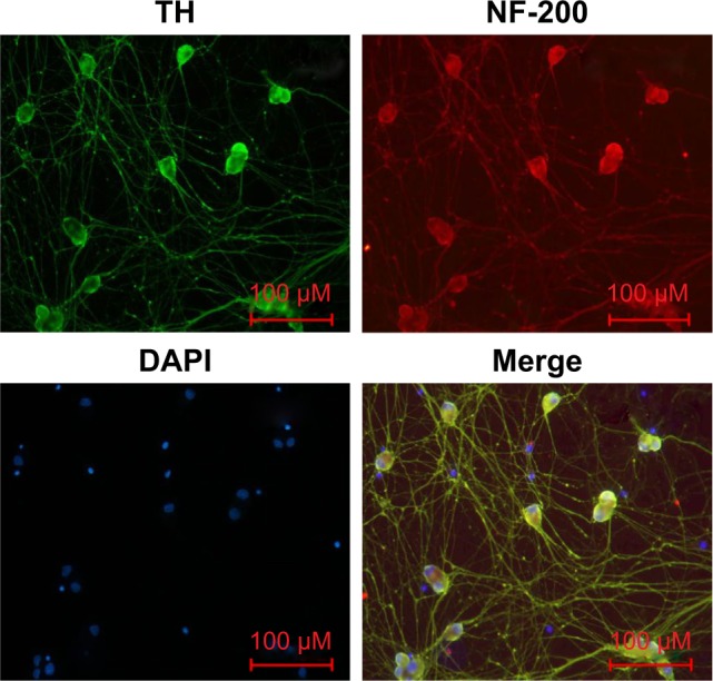
Identification of primary sympathetic neurons by immunofluorescence.
Notes: The cells derived from SCGs of newborn (1–3 days) rats were TH (green) and NF-200 (red) immunopositive. Overlayed images showed that NF-200 (red), TH, and DAPI (blue) completely overlapped (magnification ×200, bar=100 µm).
Abbreviations: DAPI, 4′,6-diamidino-2-phenylindole; NF-200, neurofilament-200; TH, tyrosine hydroxylase.