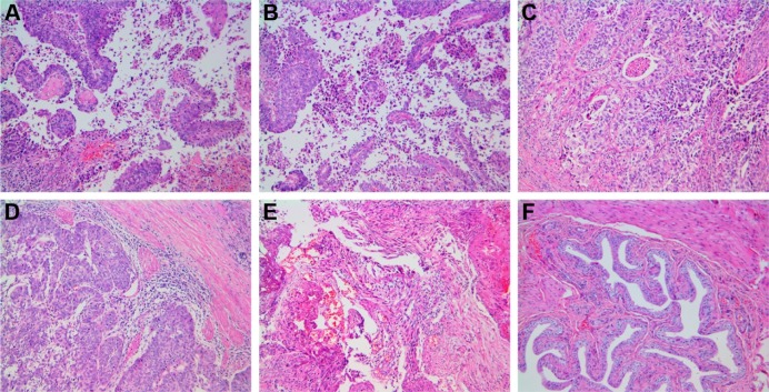
Figure 7.
Histopathology.
Notes: (A) Control; (B) LK; (C) PTX; (D) LK/P; (E) PTX/P; (F) LK/PTX/P. Disorganized cells, pathological mitotic figure, and papillary hyperplasia are observed in local or multiple regions in the Control group; the cell abnormality in LK and PTX groups is lower than that of the Control group; the cell abnormality in the LK/P and PTX/P groups is lower than that of the LK and PTX groups; and the cell abnormality in the LK/PTX/P group is significantly lower than that of the LK/P and PTX/P groups. Hematoxylin-Eosin staining, magnification ×100.
Abbreviations: LK, lumbrokinase; P, PEG-b-(PELG-g-(PZLL-r-PLL)); PEG-b-(PELG-g-(PZLL-r-PLL)), poly(ethylene glycol)-b-(poly(ethylenediamine l-glutamate)-g-poly(ε-benzyoxycarbonyl-l-lysine)-r-poly(l-lysine)); PTX, paclitaxel.

