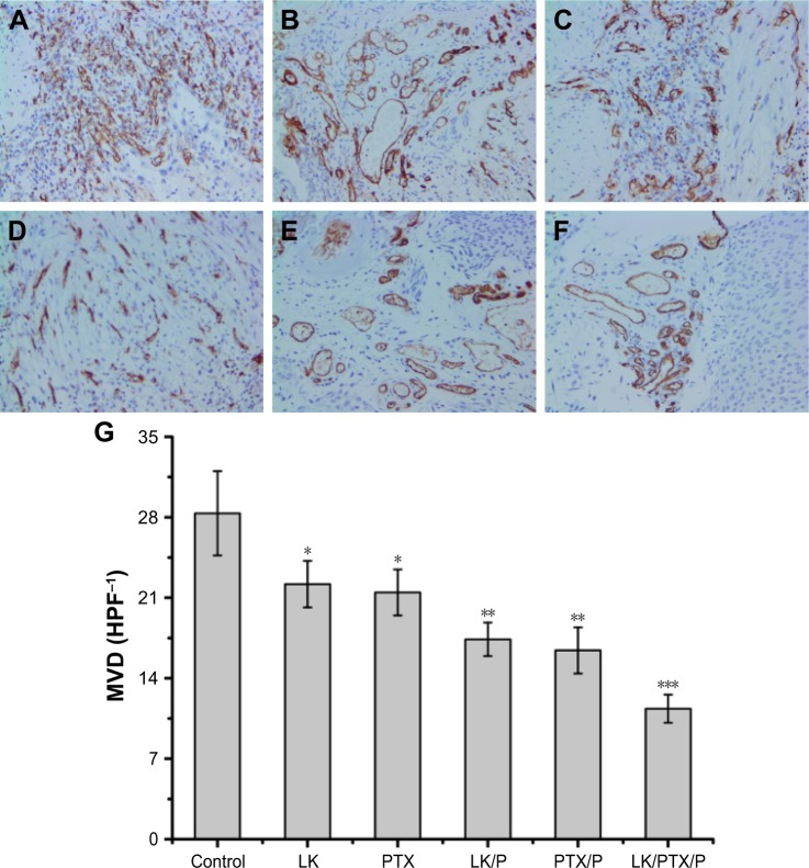
Figure 8.
CD34 expression and MVD in the rat bladder cancer tissue.
Notes: (A) Control; (B) LK; (C) PTX; (D) LK/P; (E) PTX/P; (F) LK/PTX/P; (G) MVD. Immunohistochemical staining, magnification ×200. Data are expressed as the mean ± SD, n=10. *p<0.01 vs Control, **p<0.05 vs LK or PTX, ***p<0.01 vs LK/P or PTX/P.
Abbreviations: HPF, high-power field in an optical microscope; LK, lumbrokinase; MVD, microvessel density; P, PEG-b-(PELG-g-(PZLL-r-PLL)); PEG-b-(PELG-g-(PZLL-r-PLL)), poly(ethylene glycol)-b-(poly(ethylenediamine l-glutamate)-g-poly(ε-benzyoxycarbonyl-l-lysine)-r-poly(l-Lysine)); PTX, paclitaxel.

