Figure 7.
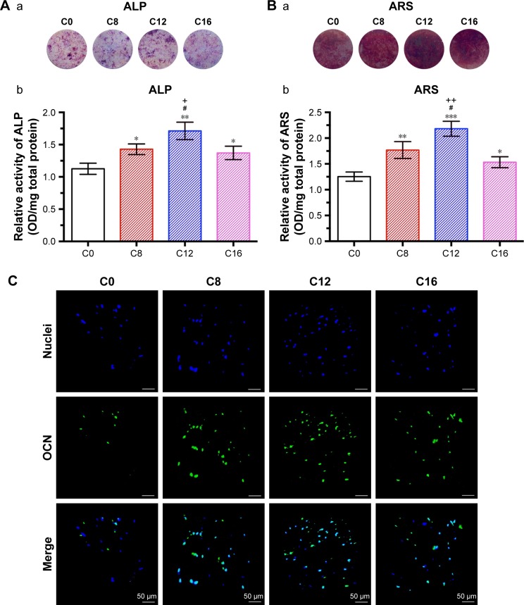
Osteogenic differentiation of rBMSCs on various coatings.
Notes: (A) ALP staining of rBMSCs cultured on different coatings for 7 days (a) and corresponding ALP activity determined with quantitative assay (b). (B) ARS staining of rBMSCs incubated on different coatings for 21 days for calcium deposition detection (a) and corresponding quantitative analysis (b). (C) The immunofluorescence assay for the expression of OCN of rBMSCs on various coatings after 7 days of incubation. Blue color, nuclei of rBMSCs stained with DAPI; green color, labeled OCN protein. “Merge” represent the merged images of nuclei and OCN. *P<0.05, **P<0.01, ***P<0.001 when compared with C0; #P<0.05 when compared with C8; +P<0.05, ++P<0.01 when compared with C16. C0, HA coating; C8–C16, differently CHA coatings.
Abbreviations: ALP, alkaline phosphatase; ARS, alizarin red solution; CHA, carbonated hydroxyapatite; HA, hydroxyapatite; OCN, osteocalcin; OD, optical density; rBMSCs, rat bone-marrow-derived mesenchymal stem cells.

