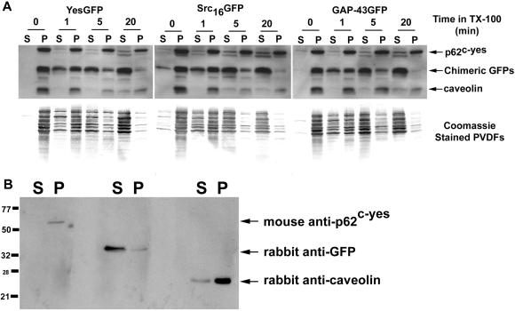
Figure 8.
Solubilization kinetics of acylated GFPs, caveolin, and p62c-yes. COS-7 cells expressing various acylated and nonacylated GFP chimeras were separated into 4°C TX-100-soluble and -resistant fractions by incubating cell monolayers with 1% TX-100 for 0–20 min. The 1% TX-100 fractions containing solubilized total cellular proteins were rapidly removed from the tissue culture dishes and the detergent-resistant matrices solubilized with a 1× lysis buffer solution. These separated fractions were immunoblotted with rabbit anti-GFP (1:2000), anti-caveolin (1:4000), and mouse anti-Yes (1:1000) antibodies. (A) Immunoblots showing relative distributions of acylated GFPs, caveolin, and p62c-yes between TX-100-soluble and -resistant fractions. Markers, right, point to the position of migration of the indicated proteins. Below each immunoblot is a corresponding Coomassie-stained PVDF membrane showing the kinetic distribution of total cellular protein between TX-100-soluble and -resistant fractions. (B) Immunoblot showing the specificity of the three antibodies used in this analysis. YesGFP is being detected in the anti-GFP blot.

