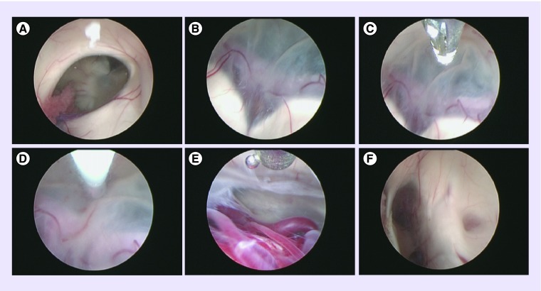
Figure 3. . Endoscopic third ventriculostomy in a child with a tectal plate/aqueductal glioma.
(A) Right foramen of Monro. (B) Floor of the third ventricle and the bifurcation of basilar artery. (C) Tip of the balloon approaching the floor of the third ventricle. (D) Ventrioculostomy using balloon. (E) Pontine perforators visible through the floor of the third ventricle. (F) Thickened tectal plate with stenosed aqueduct (on the right of the image) and pineal recess (on the left).

