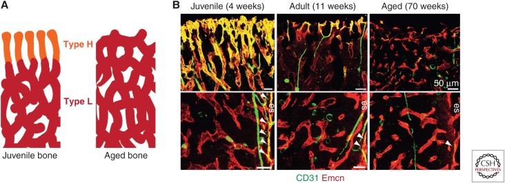
Figure 3.
Changes in bone vasculature with age. (A) Schematic diagram of changes in endothelial cell (EC) subpopulations with age, where type H vessels (orange) are gradually replaced with type L (red) vessels over time. (B) Overview fluorescent images of changes in metaphyseal (top panels) and diaphyseal (bottom panels) vessels with age. Note the age-dependent decline in type H ECs of the metaphysis and endosteum (es, arrowheads). (Image from Kusumbe et al. 2014; reprinted, with permission, from Nature Publishing Group © 2017.)

