Figure 4.
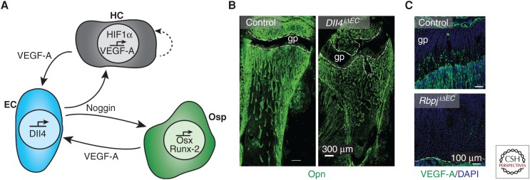
Notch signaling in the coupling of angiogenesis and osteogenesis. (A) Schematic diagram of the interaction between hypertrophic chondrocytes (HCs, gray), endothelial cells (ECs, blue), and osteoprogenitors (Osp, green). The hypoxic environment surrounding the HCs leads to accumulation of hypoxia-inducible factor (HIF)1α and subsequent expression of vascular endothelial growth factor A (VEGF-A). The VEGF-A acts as an autocrine signal for the HC as well as a paracrine signal for the neighboring EC. This induces Delta-like ligand 4 (Dll4) expression in the ECs and activates Notch signaling. Noggin is released and acts on both the HC and the Osp. Noggin promotes the generation of osteoblast lineage cells by inducing expression of Osx and Runx-2, thereby facilitating bone formation. (B) Osteopontin (Opn, green) immunostaining in 4-week-old mice lacking endothelial Dll4 (Dll4iΔEC), which display malformed bone and growth plate (gp, dashed lines). (C) Strongly reduced VEGF-A immunostaining (green) in the gp of P28 mice lacking endothelial RBP-Jκ (RbpjiΔEC), a transcription factor mediating Notch-dependent gene expression. (Fluorescent images from Ramasamy et al. 2014; reprinted, with permission, from Nature Publishing Group © 2014.)

