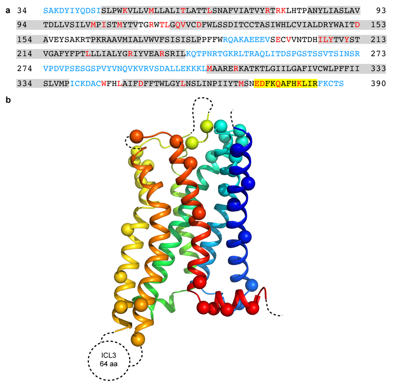
Extended Data Figure 4. Modelling quality of the 5-HT1BR structure.
a, Amino acid sequence of 5-HT1BR construct used in the cryo-EM structure determination. Residues are colored according to how they have been modelled: black, good density allows the side chain to be modelled; red, limited density for the side chain present and therefore the side chain has been truncated to Cβ; blue, no density observed and therefore the residue was not modelled. Regions highlighted in grey represent the transmembrane α-helices and amphipathic helix 8 is highlighted in yellow. b, Model of 5-HT1BR showing the Cα positions of amino acid residues with poor density (spheres) and regions unmodelled (dotted lines).

