Figure 1.

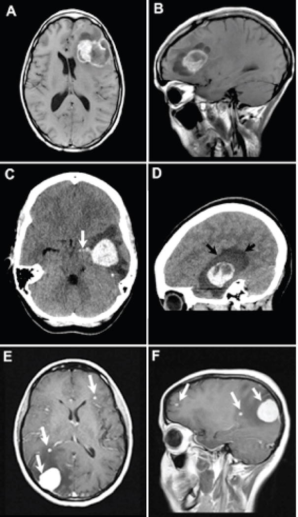
Patient 1: Axial (A) T1-weighted contrast-enhanced and sagittal (B) T1-weighted pre-contrast, demonstrate a cystic and solid metastasis within the left frontal lobe resulting in mid line shift. The metastasis contains T1 iso to hyperintense material that are iso to hyperintense on the T2-weighted images consistent with blood products (arrows). Sagittal contrast enhanced T1-weighted images depict the solid enhancing components surrounded by cystic parts of the metastasis. Patient 2: Axial (C) and sagittal (D) reformatted CT images demonstrate a hyperdense hemorrhagic metastasis within the left temporal lobe. Rupture into ventricular system with blood extending into temporal horn of the lateral ventricle [white arrow image (C)]. Edema surrounding the metastasis dark shade of gray designated by black arrows on image (D). Patient 3: Axial (E) and sagittal (F) T1-weighted contrast enhanced images reveal several enhancing metastases (white arrows).
