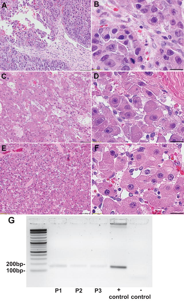
Figure 2.

H&E of resected brain metastasis at 10x (A,C,E) and 60x (B,D,F) magnification from Patient 1 (A,B) and Patient 2 (C,D) and Patient 3 (E,F), demonstrating well-differentiated tumor cells with abundant eosinophilic cytoplasm and prominent nucleoli. Fibrous bands, classic for FLHCC are demonstrated in Patient 2 (D, top right corner) and were absent in Patients 1 and 3. Scale bars 100 microns (A,C,E) and 20 microns (B,D,F). RT-PCR agarose gel electrophoresis for the DNAJB1-PRKACA fusion mRNA, is positive for a 148bp amplicon in brain metastases from Patient 1 (lane 2), Patient 2 (lane 3), Patient 3 (lane 4), positive control for DNAJB1-PRKACA (lane 5) and a negative control (lane 6). Lanes are shown from left to right with a 10Kb ladder (lane 1) (G).
