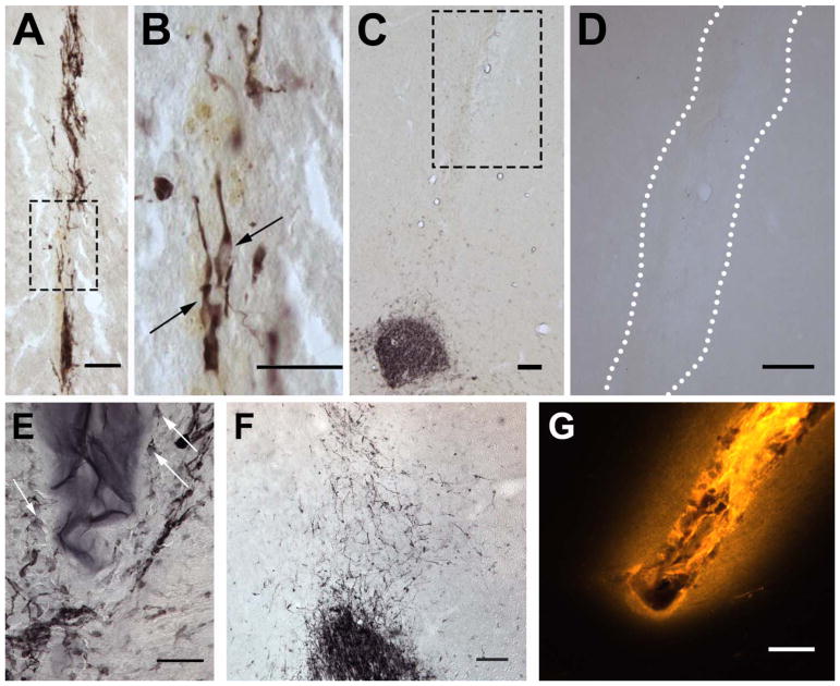
Fig 2.
Detection of neuroblasts in brain tissue by doublecortin (DCX) immunohistochemistry. Neuroblast found inside the E2Ten-C PA track (A) show elongated morphology (B) typical of migrating cells. (C) No redirected cell migration was observed with E2Ten-C PA injection (white outline) when the injection track does not reach the RMS, evident by lack of DCX+ cells along the PA track. (D) When E2Ten-C PA was injected at 1 wt% concentration cells surrounded the polymer (arrows) but none infiltrated the biomaterial due to the high density. (E) When non-polymerizing Ten-C peptide was injected cells migrated out from the RMS a short distance in a random and dispersed fashion. (F) Fluorescent image showing diffusion of PA in tissue using a fluorescent TAMRA derived molecule covalently attached to the PA. Scale bars in (A–B) and (D–F) = 50 μm. Scale bar in (C) and (G) = 100 μm.

